Brain Activity in Adolescent Major Depressive Disorder Before and After Fluoxetine Treatment
by Tao R, Calley CS, Hart J, Mayes TL, Nakonezny PA, Lu H, Kennard BD, Tamminga CA, and Emslie GJ.
American Journal of Psychiatry 2012 169 :381 –388.
Objective: Major depression in adolescents is a significant public health concern because of its frequency and severity. To examine the neurobiological basis of depression in this population, the authors studied functional activation characteristics of the brain before and after antidepressant treatment in antidepressant naive depressed adolescents and healthy comparison subjects.
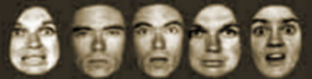
Method: Depressed [N=19] and healthy [N=21] adolescents, ages 11 to 18 years, underwent functional MRI assessment while viewing fearful and neutral facial expressions at baseline and again 8 weeks later. The depressed adolescents received 8 weeks of open-label fluoxetine treatment after their baseline scan.
Results: Voxel-wise whole brain analyses showed that depressed youths have exaggerated brain activation compared with healthy comparison subjects in multiple regions, including the frontal, temporal, and limbic cortices. The 8 weeks of fluoxetine treatment normalized most of these regions of hyperactivity in the depressed group. Region-of-interest analyses of the areas involved in emotion processing indicated that before treatment, depressed youths had significantly greater activations to fearful relative to neutral facial expressions than did healthy comparison subjects in the amygdala, orbitofrontal cortex, and subgenual anterior cingulate cortex bilaterally. Fluoxetine treatment decreased activations in all three regions, as compared with the repeat scans of healthy comparison subjects.
Conclusions: While effective treatments are available, the impact of depression and its treatment on the brain in adolescents is understudied. This study confirms increases in brain activation in untreated depressed adolescents and demonstrates reductions in these aberrant activations with treatment.
 A voxel [volumetric pixel or Volumetric Picture Element] is a volume element, representing a value on a regular grid in three dimensional space. This is analogous to a pixel, which represents 2D image data in a bitmap [which is sometimes referred to as a pixmap]. As with pixels in a bitmap, voxels themselves do not typically have their position [their coordinates] explicitly encoded along with their values. Instead, the position of a voxel is inferred based upon its position relative to other voxels[ i.e., its position in the data structure that makes up a single volumetric image]. In contrast to pixels and voxels, points and polygons are often explicitly represented by the coordinates of their vertices…
Functional magnetic resonance imaging or functional MRI (fMRI) is an MRI procedure that measures brain activity by detecting associated changes in blood flow. The primary form of fMRI uses the blood-oxygen-level-dependent (BOLD) contrast, discovered by Seiji Ogawa. This is a type of specialized brain and body scan used to map neural activity in the brain or spinal cord of humans or animals by imaging the change in blood flow (hemodynamic response) related to energy use by brain cells. Since the early 1990s, fMRI has come to dominate brain mapping research because it does not require people to undergo shots, surgery, or to ingest substances, or be exposed to radiation. The procedure is similar to MRI but uses the change in magnetization between oxygen-rich and oxygen-poor blood as its basic measure. This measure is frequently corrupted by noise from various sources and hence statistical procedures are used to extract the underlying signal. The resulting brain activation can be presented graphically by color-coding the strength of activation across the brain or the specific region studied. The technique can localize activity to within millimeters but, using standard techniques, no better than within a window of a few seconds…
Whole Brain Analyses:
At baseline, the depression group showed significantly greater activations relative to the healthy comparison group for the fearful > neutral contrast in the regions of the left and right frontal lobe, temporal lobe, putamen, insula, and cingulate gyrus and in the right amygdala, right hippocampus, and right occipital cortex [p values, <0.001], although none of the differences reached a p value of 0.05 after false discovery rate correction for multiple testing. At week 8, the depression group had greater activation than the comparison group [in a single five-voxel cluster] only at the left superior and middle frontal gyrus [MNI coordinates: x=–25, y=44, z=4; z=3.66, t=4.16, uncorrected p<0.001; false discovery rate corrected p=0.76]…
 My point is not to devalue the work these people are doing. Reading this study, I had the feeling that they are trying hard to correlate clinical states or responses to medications with changes they can actually measure in the brain. There’s a ton of technology involved and a graduate course or two in the math they’re using to get an output. Something about being a teenager rolled into a MRI looking at those pictures seems really kind of far-fetched to me – the stuff of bad dreams. But that aside, using subtle changes in brain blood flow to localize things happening in the brain is pretty damned interesting. This is way, way off-Broadway stuff, in my estimation, but it has the feel of something that may be useful way, way down the road – but not right now or in the near future. I was unconvinced that the conclusion "This study confirms increases in brain activation in untreated depressed adolescents and demonstrates reductions in these aberrant activations with treatment" was justified.
I see some problems with this kind of research for the future. It’s virtually impossible to show the results in a way that the reader can evaluate them – even the statistical tools for multiple sampling are somewhat arbitrary and lack the precision of other places where statistical analysis is useful. This is exciting technology, and I can see why people are so taken with it, but it is still experimental at best. In this case, the senior author, Graham Emslie, is someone who has been involved in the majority of the research supporting the effectiveness of anti-depressants in youths, from Paxil Study 329 through all the studies in the recent Gibbons’ meta-analysis, and who has extensive industry connectedness. His bias in these matters is likely.
Oh, those voxels. I’d like to see what Daniel Bor http://www.danielbor.com/dilemma-weak-neuroimaging/ has to say about this study.
My take is that these study situations are as close to human reality as forced-swim studies in mice are, and the researchers see what they want to see in the data.
My estimate is that this imaging studies and those like it are about 6 degrees removed from reality.
The first degree of error is in diagnosis: How did they determine which adolescents were clinically depressed?
Second degree: How close is the study condition (MRI machine) to real life? (Could the study condition itself produce some kind of emotional reaction? I hate being in one of those machines myself.)
Third degree: How valid is the interpretation of a global emotional state from momentary reactions to stupid pictures?
Fourth degree: Could the statistical analysis methodology of the voxels be influenced by the interests and expectations of the researchers? (Daniel Bors, white courtesy telephone!)
Fifth degree: Could the interpretation of the charts resulting from statistical manipulation be influenced by the interests and expectations of the researchers? (The new phrenology.)
Sixth degree: Could the interpretation of statistical significance of the data gathered from the two (possibly misdiagnosed) groups be influenced by the interests and expectations of the researchers? (Same old, same old.)
Given Graham Emslie is involved, you know what this team wanted to find. Everything coming out of this study is suspect.
This is the kind of tangentially relevant research that I call Ganser-ing. Ganser’s Syndrome is known as the syndrome of approximate answers. In research terms, this study typifies the syndrome of approximate questions: the depressed patients were challenged with fearful or neutral faces. Did the authors not have any sad or glad faces to use instead? Why study fear when you could study hedonic responsiveness? Why study a tangential aspect of depression when you could study a central aspect of the condition?
This tells us that the authors didn’t think carefully about their research design, but rather used an off-the-shelf experimental procedure. No wonder they obtained only an approximate answer.
I invite everyone to read the comments by other neuroscientists on Daniel Bor’s article on neuroimaging: http://www.danielbor.com/dilemma-weak-neuroimaging/
Marc Pelletier of Case Western says: “In clinical fMRI, it seems to me that there is no consensus about the thresholds and the methods to be used to objectify a significant group difference for brain activations.”
In any task, the entire brain is active. Determining a significant circuit is like reading tea leaves.
What a fine tip from Altostrata [the post and comments] on Daniel Bor’s blog: http://www.danielbor.com/dilemma-weak-neuroimaging/. It’s a nesting place for neuroimaging gurus. I invited them to weigh in on this article. I hope they’ll consider taking it on. It needs taking on…